 치과소개
치과소개 자연치아살리기
 수면마취
수면마취 임플란트
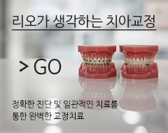
임플란트 치아교정
치아교정 심미치료
심미치료 일반치료
일반치료 사랑니발치
사랑니발치
 진료철학
진료철학 의료진 소개
 둘러보기
둘러보기 첨단장비
첨단장비 소독멸균
소독멸균 리오기공소
리오기공소 무통마취
무통마취 오시는길
오시는길 의료진소개
의료진소개 오시는길
오시는길
 리오가 생각하는 치아
 재근관치료
재근관치료 엠도게인
엠도게인 리오가 생각하는 치아
 엠도게인
엠도게인
 수면마취란?
 수면 임플란트
 수면 사랑니발치
 주의사항
주의사항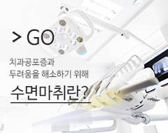 수면마취란?
 수면임플란트
 리오가생각하는임플란트
 UV임플란트
 전체임플란트
 즉시임플란트
 비절개 임플란트
 네비게이션 임플란트
 임플란트 틀니
 임플란트 재수술
 전신질환 임플란트
 임플란트 시술후 관리
 리오가생각하는임플란트
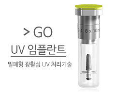 UV임플란트


 심미치료란?
 라미네이트
라미네이트 올세라믹
올세라믹 잇몸성형
잇몸성형 치아미백
치아미백 잇몸미백
잇몸미백 심미치료란?
 라미네이트
라미네이트
 충치치료
충치치료 신경치료
신경치료 치주치료
치주치료 스케일링
스케일링 시린이
시린이 예방치료
예방치료 틀니
틀니 턱관절 치료
 수면 사랑니발치
 충치치료
충치치료 신경치료
신경치료
 리오가생각하는사랑니발치
 사랑니발치
사랑니발치 수면사랑니발치
 주의사항
주의사항 리오가 생각하는 사랑니발치
 수면사랑니발치
Membrane interaction (Figure 2B). As with HIV, a hydrophobic cavity fo…
페이지 정보
작성자 Maynard 댓글 0건 조회 135회 작성일 24-04-09 09:38본문
Membrane interaction (Figure 2B). As with HIV, a hydrophobic cavity formed by the side chains of helices h1 to h4 is observed in the FIV p15 protein (Figure 2B). Our docking studies demonstrated that a myristoyl group can be placed in this cavity with its carboxyl end in the vicinity of the Nterminus of p15, providing the local adjustment of residues from helices h1, h2 and h4 (Trp9, Phe35, Ile39, n-Phenylpiperazine-1-carboxamide Ile53, and Phe90, Figure 2C). These residues are located at the same positions as the residues interacting with the N-terminal myristoyl of the HIV MA (Ser9, Ile34, Ser38, Leu51 and Leu85, respectively [21]). The estimated binding free energy of the myristoyl group is similar for FIV and HIV (-2.9 kcal/mol and -3.7 kcal/mol, respectively). One striking feature, when the myristoyl group is inserted in the hydrophobic pocket of FIV p15, is the predicted motion of Trp9 and Ile53, which rotate from their initial position (Figure 2C). Of interest, our docking experiments revealed a second putative binding site for the myristoyl group that wraps around the first turn of helix h1 (Figure 2D) in a groove located at the surface of the protein (Figure 2E). This location involves the interaction of themyristoyl group with residues Trp9, Ala12, Arg15, Glu55 and Leu95 of helix h1 and loops l2 and l4. FIV p15, despite having a low sequence similarity with HIV, EIAV and SIV MA (18 , 17 and 15 sequence identities, respectively) is strikingly similar in structure to these lentiviral matrix PubMed ID:https://www.ncbi.nlm.nih.gov/pubmed/20618955 proteins and can be superposed to each with a root mean square deviation (RMSD) of 1.9 ? 2.5 ? and 2.1 ?between C pairs, respectively (Table 2). However, the superimposition of the lentiviral MA structure reveals a major difference around the C-terminal end Fmoc-D-Asp-OtBu of MA (Figure 3). The C-terminal end of FIV p15 is mainly unstructured and extended (Figures 2 and 3), whereas a long helix h5 is observed in HIV p17 (Figure 3A), and a short -hairpin is reported in SIV MA (Figure 3B). The C-terminal end of EIAV p15 is not visible in the electron density maps and is absent from itsTable 2 Structural comparisons with other lentiviral matrix proteinsProtein EIAV MA HIV MA SIV MAPDB ID 1HEK 1HIW 1ECWRMSD1 2.5 ?1.9 ?2.1 ?N2 align 99 101seq3 17 18Root Mean Square Deviation calculated between C-atoms of matched residues. 2 Number of aligned residues after structure superimposition. 3 Sequence identity.Serri e et al. Retrovirology 2013, 10:64 http://www.retrovirology.com/content/10/1/Page 6 ofFigure 3 Structural comparison of four lentiviral matrix protein. The superimposition of the matrix proteins of FIV (red) with (A) HIV (PDB ID: 1HIW, blue), (B) SIV (PDB ID: 1ECW, green) and (C) EIAV (PDB ID: 1HEK, yellow) matrix proteins. The N- and C-termini are indicated, and the helices are numbered h1 to h5 as in Figure 2A. Black arrows highlight the structural differences at the C-terminal end of the matrix proteins.structure (Figure 3C). Another difference is the presence of longer loops in FIV as compared with EIAV, HIV and SIV because of insertions in the FIV sequence (Figure 4). As these loops are important to the stabilization of HIV and SIV trimers, the longer PubMed ID:https://www.ncbi.nlm.nih.gov/pubmed/11834444 loops may be related to the differences observed in the FIV p15 oligomerization. There are 6 conserved residues between the proteins (Figure 2-dioxaborolane 3-Fluoro-5-iodobromobenzene Cyclobutylboronic acid 4, black squares), most of which are within the helical sections of the molecule, and they may be responsible for maintaining the characteristic matrix architecture. Finally, we can observe in the.
- 이전글Amoxicilín na Slovensku Amoxicilín kde kúpiť 24.04.09
- 다음글Glimepirid skusenosti Glimepirid Prešov 24.04.09
댓글목록
등록된 댓글이 없습니다.